|
|
An ultrasound is a method of obtaining images of your internal organs by directing inaudible sound waves into your body with a hand-held instrument called a transducer. As the sound waves make contact with blood, tissue, and bone, portions of the sound waves echo back to the transducer. These echoes are then analyzed by a computer to produce an image of the organs on a TV-like screen. There are different types of ultrasound studies. Sometimes ultrasound is used in combination with another procedure, such as a biopsy. The exact study you are scheduled for will vary depending upon your physician’s request and the reason for the study.
A note to inpatients: If you are having this procedure done on an inpatient basis, your nurse will guide you through all preparations, and will arrange for your transportation to and from the Radiology Department.
The preparation for your ultrasound depends on the type of examination to be performed. Please check the information listed for your particular study.
When you arrive for your appointment, you will be directed to a dressing room and asked to change into an exam gown. When you are ready, a technologist will escort you to an Ultrasound Examination Room, and help you onto the scanning table. To prepare you for the exam, the technologist will rub a gel over the area of your body being studied. This gel provides better contact between the ultrasound transducer and your skin. As the study begins, the ultrasonographer will place the transducer against your skin and pass it over the area being studied.
Remember: ultrasound is a safe, painless, noninvasive procedure. No x-rays are involved.
The images acquired during this procedure will be reviewed by a radiologist who will then report the results to your physician. Your physician will discuss the results of your ultrasound at your next appointment.
| Study |
Preparation |
Time
frame |
| Abdominal
Ultrasound |
No
food or fluids for 6 hours before study. |
30
minutes |
Abdominal
and Pelvic
Ultrasound |
No
food for 6 hours before study. Drink 32 oz water
1 hour prior to study. DO NOT EMPTY BLADDER; YOUR
BLADDER MUST BE FULL. |
30
minutes |
| Pelvic
Ultrasound with Transvaginal Ultrasound if necessary |
Drink
32 oz. of fluid 1 hour prior to study. DO NOT EMPTY
BLADDER; YOUR BLADDER MUST BE FULL. |
1
hour |
| Transvaginal
Ultrasound ONLY |
No
preparation. |
30
minutes |
| Pelvic
and Transvaginal Ultrasound |
Drink
32 oz. of fluid 1 hour prior to study. DO NOT EMPTY
BLADDER; YOUR BLADDER MUST BE FULL. |
1
hour |
| Fetal/Level
I Ultrasound |
Drink
32 oz. of fluid 1 hour prior to study. DO NOT EMPTY
BLADDER; YOUR BLADDER MUST BE FULL. |
30
minutes |
| Carotid
Doppler |
No
preparation. |
30
minutes |
| Renal
Ultrasound |
No
preparation. |
30
minutes |
| Aortic
Ultrasound |
No
preparation. |
30
minutes |
| Breast
Ultrasound |
Must
have recent mammogram. If done elsewhere,
please bring film with you. |
30
minutes |
| Thyroid
Ultrasound |
No
preparation. |
30
minutes |
| Testicular
or Scrotal Ultrasound |
No
preparation. |
30
minutes |
| Lower
Extremity Venous or Arterial Ultrasound |
No
preparation. |
30
minutes |
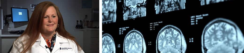
| Head
Ultrasound |
No
preparation. |
30
minutes |
| Prostate
or Transrectal Ultrasound |
Fleet enema
1 hour before study; instructions on kit. |
30
minutes |
| Biopsies |
No
food or fluids for 6 hours before study. Blood tests
may be needed. Must be accompanied by driver; patient
will not be allowed to drive home. |
1
hour; patient observed for an additional hour |
| Chest/Pleural
Effusion |
Must
have a chest x-ray taken within one week of study.
If done elsewhere, please bring film with you. |
30
minutes |
|
Back to Top
You should report to The Reading Hospital and Medical Center (location specified by your physician) at the time indicated. After you are registered, you will be escorted to a special nursing unit where you will be prepared for the procedure. The arteriogram will be performed in the Hospital's Interventional Radiology Suite. You will be taken to this section of the Radiology Department on a stretcher. Following the procedure, you will be taken to your assigned room to relax.
If you have diabetes requiring insulin, it may be necessary to make some adjustments. Please contact your physician. We also recommend that you leave jewelry, hair pins, contact lenses, dentures, etc., at home.
The arteriogram will be performed approximately two hours after your arrival at the Hospital. This early arrival is necessary for the Nursing staff to complete the preparations outlined here. First, the skin near the site where a catheter will later be inserted needs to be shaved. In most cases, this site is the groin (at the top of the leg). Less frequently, the upper arm may be used. (The catheter will actually be inserted later, once you are taken to the Radiology Department.) Next, the nurse will place an intravenous tube in your arm in order to give you additional fluids. Before the study, you will change into a patient gown, and, if you did not leave them at home, all jewelry, hair pins, dentures, etc., will be removed. The nurse will then give you an injection to relax you. This may make you drowsy. You will then be transported to the Radiology Department.
The Interventional Radiology room is similar to other x-ray rooms except that it contains more equipment. After you have been helped onto the table, preliminary x-rays will be taken. A technologist will then scrub your groin or arm with an antiseptic solution, and will drape the area with sterile towels. There are usually one or two physicians and two or three technologists present during the study. Because you will be awake during the study, you will receive local anesthesia so that you remain comfortable. This medication may burn when first injected. However, within a few seconds, the area will become numb. Thereafter, you will feel only mild pressure when the catheter is inserted and manipulated. When the catheter is introduced into the blood vessel, the room will be darkened, and a picture will appear on a small overhead screen. The physician is able to see the catheter on this screen, and is able to tell when it is correctly positioned. When this has been accomplished, the physician will inject the dye. For many people, the injection causes a hot flushing feeling and the sensation that they have urinated. Please do not become alarmed. These sensations last only a few seconds. During the introduction of the dye, x-ray films are taken. Upon the completion of the study, the physician will remove the catheter, and apply pressure to the insertion site to stop any oozing. Sometimes a special stitch or patch may be applied to the site. A dressing will be secured to the puncture site. The entire procedure takes between one and two-and-one-half hours, depending upon the complexity of the examination. For most people, the greatest discomfort is mild backache from lying in the same position for that long of a time period.
You will be taken back to your assigned bed, where you will remain between six and nine hours. The nurses will examine your dressing, and monitor your pulse and blood pressure carefully. Because you will remain in bed, it may be necessary for you to use the bedpan to void or move your bowels. You may, however, perform any activities that you can safely do in bed: eat, watch TV, read, and have visitors. At the end of the time, you may resume your usual activity. The x-ray films will be reviewed by the radiologist. This physician will then report the results to your doctor, who will discuss them with you at your next appointment.
Back to Top
|